Utilizing Dino-lite Digital Microscope for Failure Analysis of PCB Boards
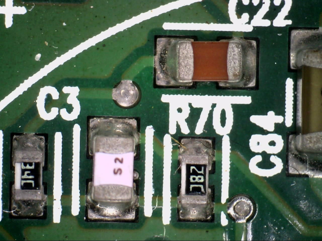
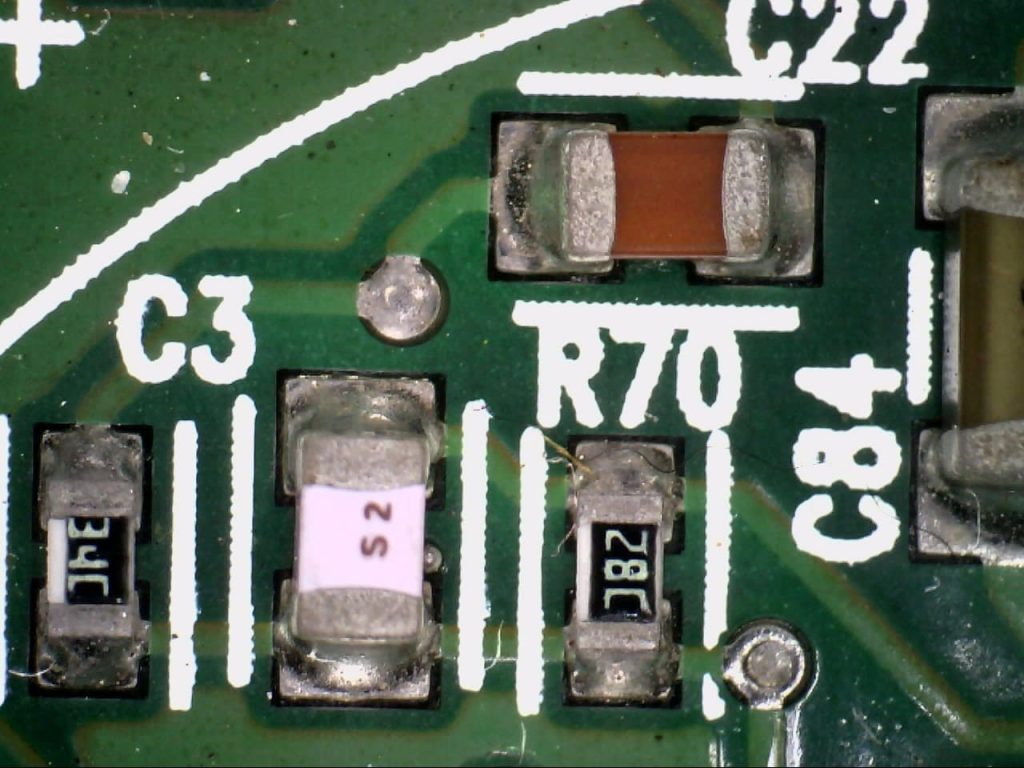
Failure analysis is the process of collecting and analyzing data to determine the cause of a failure, often with the goal of determining corrective actions or liability. The failure analysis process relies on collecting failed components for subsequent examination of the cause or causes of failure using a wide array of methods, especially microscopy. Optical microscopy may be one of the most popular and preferred testing methods used for detecting faults, defects and problems associated with soldering and assembly. Many customers choose optical microscopy because of its speed and accuracy. Microscopy testing can verify improper construction, which can lead to stresses that can expose flaws at certain cross sections. Dino-Lite has been used increasingly for failure analysis in the microelectronics industry, especially for printed circuit boards (PCBs). Digital microscopes are practical to use and allow an efficient workflow for inspection.
Dino-Lite Digital Microscopes in PCB Failure Analysis
Printed circuit boards (PCBs) are the intricate lifeblood of modern electronics. When a PCB fails, malfunctions can range from minor inconveniences to complete device failure. Identifying the root cause of these failures is crucial for improving product quality and ensuring device reliability. This is where digital microscopes step in, offering a powerful tool for PCB failure analysis. Traditionally, PCB failure analysis relied on visual inspection using magnifying glasses or low-powered microscopes. While these methods provide a basic view, they often fall short when dealing with highly miniaturized components and intricate defects. Additionally, documentation of these observations can be challenging and subjective.
Dino-Lite Digital Microscopes: A High-Resolution Advantage
Dino-Lite offers a significant leap forward in PCB failure analysis. Their key advantages include:
- High Magnification: Ranging from 20x to 200x or even higher, digital microscopes allow for detailed observation of solder joints, component leads, and PCB surface features. This magnification helps identify subtle defects like cracks, voids, delamination (layer separation), and improper solder formation.
- Digital Imaging and Documentation: Captured high-resolution images and videos provide objective and sharable records of the failure site. This facilitates collaboration between engineers and failure analysts, allowing for more efficient troubleshooting.
- Enhanced Lighting: Adjustable LED illumination ensures optimal lighting for examining different areas of the PCB. Specific lighting modes can further reveal details, such as the presence of contamination or corrosion.
- Measurement Capabilities: Some digital microscopes offer built-in measurement tools for precise quantification of defect size, solder joint width, or component lead diameter. This data can be crucial for pinpointing the root cause of the failure.
Failure Analysis with Digital Microscopes: A Step-by-Step Approach
The use of digital microscopes in PCB failure analysis typically follows a systematic approach:
- Initial Inspection: The PCB is visually inspected to identify potential areas of concern like burn marks, discoloration, or component damage.
- High-Resolution Examination: The Dino-Lite digital microscope is used to examine these areas in detail, capturing images and videos for documentation.
- Defect Identification: Based on the magnified view and knowledge of common PCB failure modes, the specific defect is identified (e.g., solder joint crack, component overheating).
- Measurement and Analysis: Measurements are taken of the defect size and surrounding features. This data helps determine the potential cause and severity of the failure.
- Root Cause Determination: By analyzing the findings, the root cause of the failure can be identified, such as manufacturing process issues, component selection errors, or design flaws.
The Benefits of Dino-Lite
The use of digital microscopes in PCB failure analysis offers several advantages:
- Improved Accuracy: High-resolution magnification allows for more precise identification of failure modes, leading to more effective corrective actions.
- Enhanced Collaboration: Digital documentation facilitates communication between engineers and failure analysts, accelerating the troubleshooting process.
- Data-Driven Analysis: Measurements obtained through the microscope provide valuable data for failure analysis reports and future design improvements.
- Preventive Maintenance: By identifying recurring failure modes, manufacturers can implement preventive measures to improve overall PCB quality and reliability.
In Conclusion:
Digital microscopes are transforming PCB failure analysis. Their high magnification, digital imaging capabilities, and potential for AI integration make them an indispensable tool for identifying and understanding the root causes of PCB failures. By leveraging this technology, manufacturers can ensure the reliability of their electronic products and prevent future malfunctions.
Uses of Dino-lite Digital Microscope in Textile - Fabric Inspection Industry
Textile - Fabric Inspection Industry
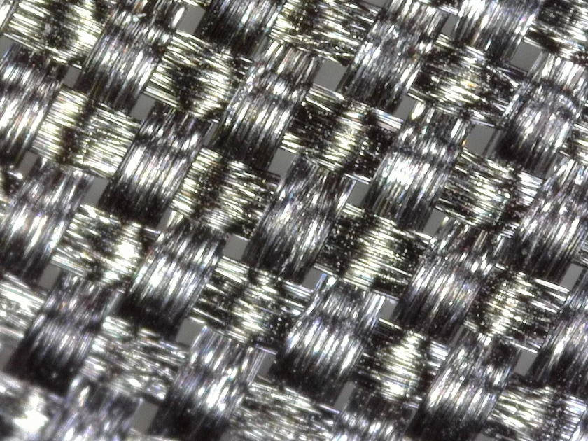
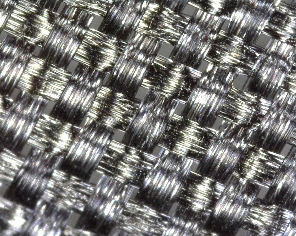 Dino-Lite digital microscope cameras allow Textile and Composite industry professionals the ability to see thread counts, contaminate section, and loom sewing monitoring with the ability to capture images and video with the included software.
How To Connect Wf-20 To Dino-lite AF Series Microscopes?
What is the WF-20?
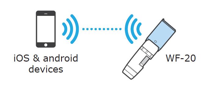
The WF-20 is a Wi-Fi streamer, as well as a wireless router, to be used with Dino-Lite Edge AF series digital microscopes for viewing and sharing observations wirelessly. The WF-20 is ideal for on-field use as it comes with high quality image transmission and a long battery life. When your AF series Dino-Lite digital microscope is connected to the WF-20, it becomes a wireless digital microscope. This is also sometimes known as a Wi-Fi digital microscope.
Compatible with mobile device or computer with operating system:
- IOS 10.X or later
- Android 6.0 or later
- Windows XP (SP3)/Vista/7/8/10
Do take note that the Dino-Lite WF-20 Wi-Fi streamer is only compatible with Dino-Lite Edge AF series digital microscope.
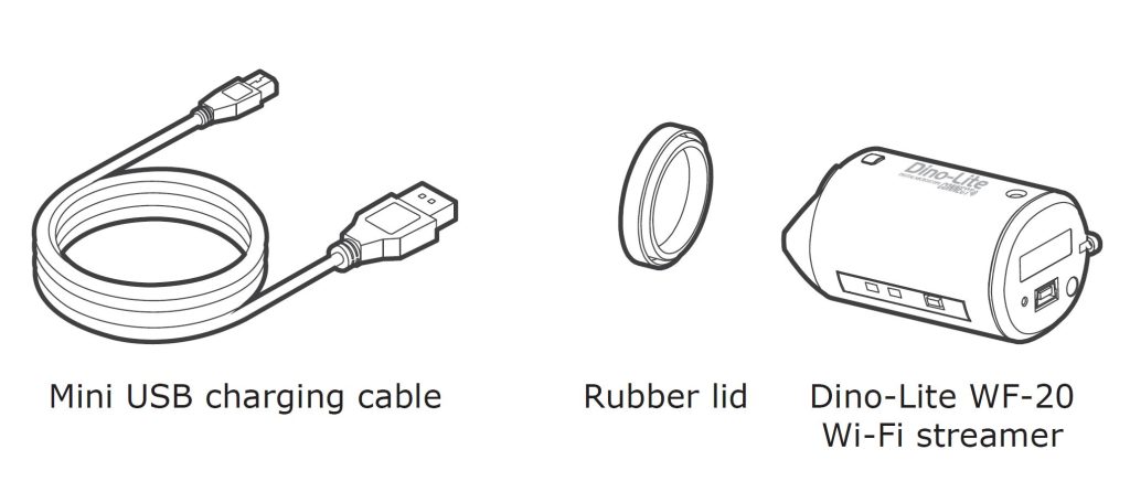
What's in the package?
WF-20 Device Overview
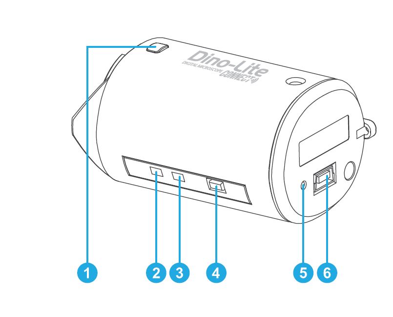
- Interface release button
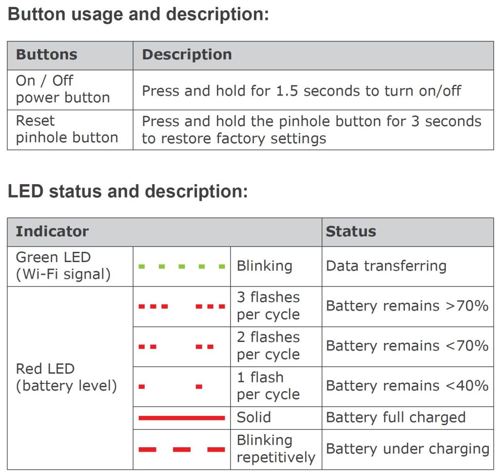
- Wi-Fi signal LED indicator
- Battery LED indicator
- On / Off power button
- Reset pinhole button
- Mini USB port (for charging purpose)
Let's get started!
Before anything, make sure you charge the WF-20 by connecting to a computer or a USB charger with the Mini USB charging cable.
1. Attach the WF-20 to Dino-Lite AF Series.
Press the two release buttons on the Dino-Lite AF series at the same time to remove the USB interface adapter.
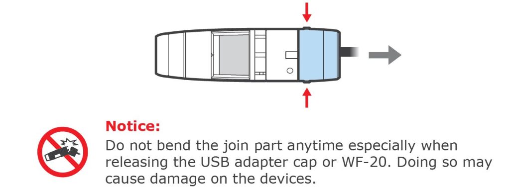
Remove the rubber lid from the WF-20. We suggest that you use the rubber lid to cover the USB adapter cap. This helps to prevent dust build-up inside the adapter.

Attach the WF-20 onto AF series with the Dino-Lite logos facing the same side.
2. Install Software
Download and install DinoConnect from the Apple App Store on iOS or Google Play store on Android.
OR
You can also install DinoCapture 2.0 which can be downloaded from https://www.dinolite.sg/downloads/software.
3. Set up the Network
Power on the Dino-Lite AF series with the WF-20 attached by pressing the power button for 1.5 seconds. The start-up process can take up to half a minute before the Dino-Lite LEDs light up. Anyway, the way to power off is also by pressing on the power button for 1.5 seconds.
On iOS/Android
- Go to settings of your iOS/Android device
- Turn on Wi-Fi.
- Select WF-20’s SSID (default: “Dino-Lite WF-20”) and input password (default: “12345678”)
- Launch DinoConnect and your Dino-Lite is ready to use!
- (Optional) When you are using multiple WF-20 nearby, be advised to change WF-20’s SSID and password in DinoConnect settings to distuingish them.
On Windows PC/laptop
- Turn on Wi-Fi of your computer, and select the network by clicking on the icon in the notification area
- Select WF-20’s SSID (default: “Dino-Lite WF-20”), and click the Connect button
- Enter the security key (default: “12345678”), and click Next to connect.
- Launch DinoCapture 2.0 and your Dino-Lite is ready to use!
Connecting to a Wireless Network
You may further setup the WF-20 as a router to build connection between your mobile and a wireless network.
- Open DinoConnect settings
- Tap “Choose a network"
- Choose a network, then enter password to connect.

Now your device is connected to the chosen network via the WF-20. The device will remember the last connected wireless network. The WF-20 can be possibly connected up to 10 mobile devices at the same time, but it is recommended to keep less than 5 connections for retaining image fluency.
You can also watch the video on how to connect to WF-20 here:
LFC is the exclusive distributor of Dino-Lite digital microscope in Singapore. Contact us if you need more information or a product demonstration. You can also visit our blog to discover more about the wireless digital microscope we carry.
How To Calibrate Your Dino-lite Digital Microscope?
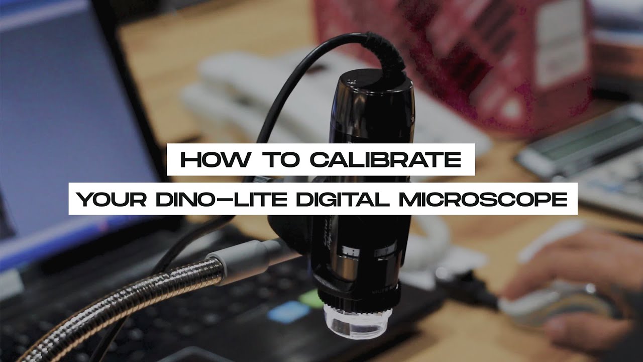
The USB version of Dino-Lite digital microscope is able to make measurements. This is done through the software DinoCapture 2.0 in Windows, and DinoXcope in Mac. In order to make measurements, you first need to calibrate your Dino-Lite.
Here are the steps on how to calibrate your Dino-Lite digital microscope:
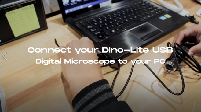
1. Connect your Dino-Lite USB digital microscope to your PC
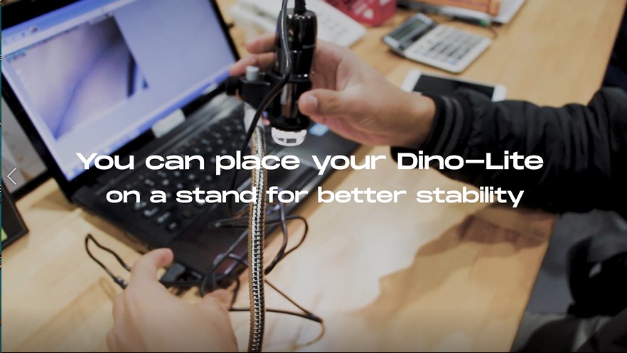
2. You can place your Dino-Lite on a stand for better stability.
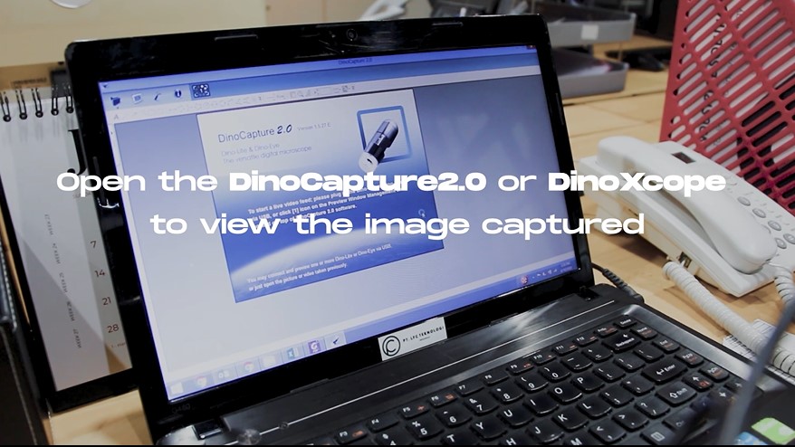
3. Open the DinoCapture2.0 or DinoXcope to view the image captured.
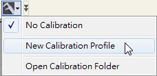
4. Click on the calibration icon on the top right hand corner and select a new calibration profile. You can name the calibration profile. For example, if you are calibrating at 200X and measuring 1mm, the profile name could be “200X – 1mm”.
5. Click “Continue Calibration” after naming the profile.
6. Place the calibration target under the microscope and focus to calibrate your Dino-Lite.
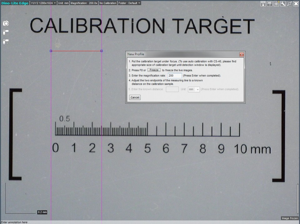
7. Click on one of the blue dots with pink guidance line to start moving to your desired location. Click again to stop. Click on the other blue dot to start setting the other end point. When the correct distance is measured, enter the known distance. In this example I am measuring a 1mm calibration target so I entered 1 into the known distance box.
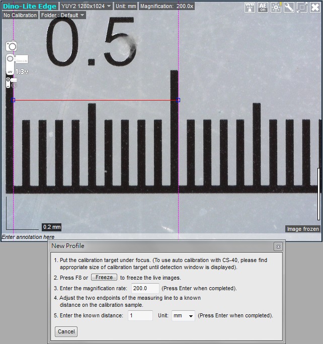
8. You have now finished setting up a new calibration profile. You are recommended to create multiple calibration profiles for different magnification levels.
For best results, it is recommended that you select a calibration profile close to the magnification you are using. For example, if the microscope is at 75X magnification and you have calibration profiles setup for 50x, 80x and 200x, it is best to use the profile setup at the closest magnification, which in this case would be 80x.
If you measure with a magnification out of the calibrated range of your profile, the results may not be accurate. For example, if you calibrate at around 50x but then measure at 200x magnification, the measurement results may be inaccurate.
If fixed magnification is required, simply calibrate at that magnification only for the most accurate results. For example, if you will be using a fixed magnification of 50x, setting up a single calibration profile at 50x will give you the most accurate results.
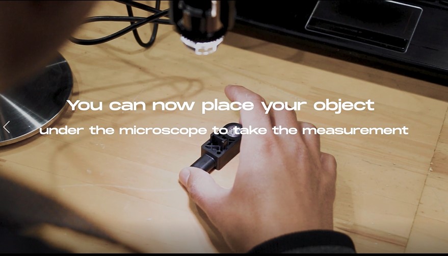
You can now place your object under the microscope to take the measurement.
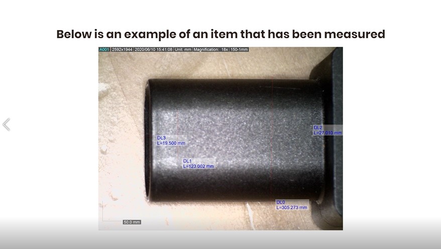
Or you can check out our video on How to calibrate your Dino-Lite Digital Microscope below :
Uses of Dino-lite Digital Microscope in Research & Development
Interactive platform for peer studying on biological and general microscopy. Dino-Lite microscope provide one of the most important platform in the field of studying biological sciences and general microscopy science. It allows students and lectures as the user to peer into the world of the cell, as well as discover the fascinating world of microscopic organisms and specimen with micron precision.
Detect Abnormal Scar Formation
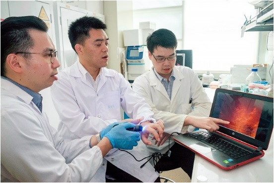
It was very hard and painful to detect skin diseases. The only tool was to perform a biopsy, where a skin tissue sample is extracted and sent for laboratory testing. The procedure is very inconvenient for patients, and an open wound also risks infections and needs suture which must be removed later.
Recently, a new technique was developed by a team led by Assistant Professor Xu Chenjie from Nanyang Technological University’s School of Chemical and Biomedical Engineering, nanoscience expert Professor Chad A. Mirkin from Northwestern University and Dr Amy S. Paller, Chair of Dermatology at Northwestern University Feinberg School of Medicine.
The new method is much more painless and allows doctor to take preventive measures earlier to help reduce the chances of serious scarring. Using new nanoparticles, the joint research team has shown in animals and human skin samples the potential to quickly and accurately predict whether a wound is likely to lead to excessive scarring as occurs in keloids and skin contractures.
Published last month in the Nature Biomedical Engineering journal, the new detection method uses thousands of nanoparticles called NanoFlares, which have DNA strands attached to their surfaces like a ball of spikes.
These nanoparticles are applied to closed wounds using a cream. After the nanoparticles have penetrated the skin cells for 24 hours, a handheld fluorescence microscope is used to look for signals given out by the nanoparticles’ interaction with target biomarkers inside the skin cells.
Using the Dino-Lite AM4115T-GFBW, when fluorescence signals are detected, they indicate abnormal scarring activity and preventive action can be taken to hopefully avoid heavier scarring.
Assistant Professor Xu Chenjie said: “When our bioengineered nanoparticles are applied on the skin, they will penetrate up to 2mm below the skin surface and enter scar cells.”
In other recently published or accepted peer-reviewed journal articles, such as in a SLAS Technology commentary, Dr Yeo further elaborates on the potential applications of NanoFlares for other skin diseases such as skin cancer, since the DNA sequences on the nanoparticles are interchangeable.
The team has filed a patent application based on this technology through NTU’s innovation and commercialisation arm, NTUitive, and will conduct a clinical trial by mid-2019.
References:
New nanoparticles help to detect serious wound
Uses of Dino-lite Digital Microscope in Educational
Over the past eight years, the Dino-Lite digital microscope has helped users around the world increase their productivity in a wide range of mundane to mission-critical applications. Due to its superior image quality, compact form factor, and ease-of-use, this product has quickly risen to a dominant position in the marketplace. From its initial success, the product line has expanded to include hundreds of models ( and accessories) to fulfill specific customer needs. As a result, the Dino-Lite microscope is now an indispensable tool in multiple market sectors, such as academics, manufacturing, quality control, healthcare, and education.
1. Art Conservation & Restoration
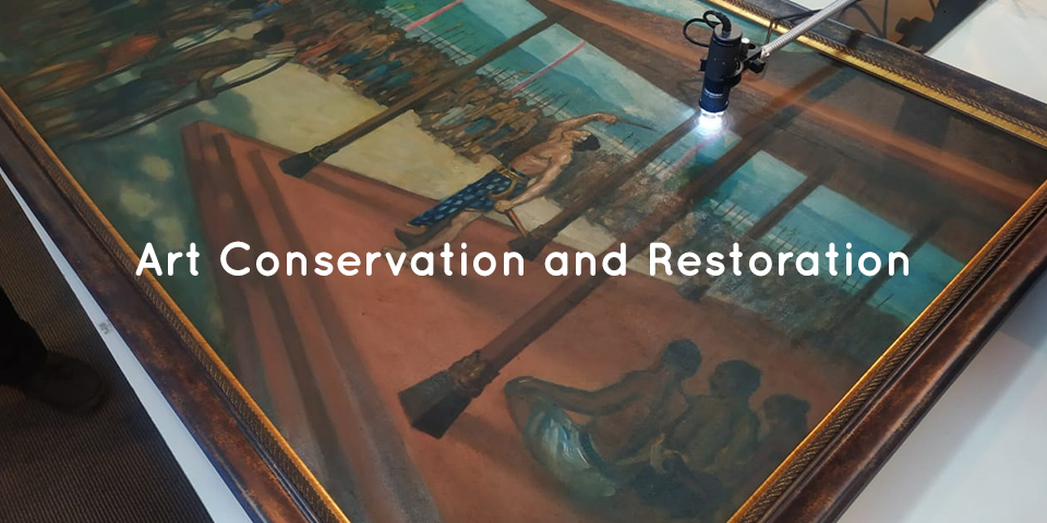
Art conservation is both an art and a science. The object of interest ranges from sculptures, ceramics, paper scroll (books, rolls, etc.), textiles and even architecture. Some of these could be performed in the lab, while others can only be done onsite. A microscope is a necessary tool in art conservation and restoration. They are used in non-destructive analysis and characterisation tests of the piece. Hence, the Dino-Lite digital microscope being small, light and portable is a great help for anyone in this field of work.
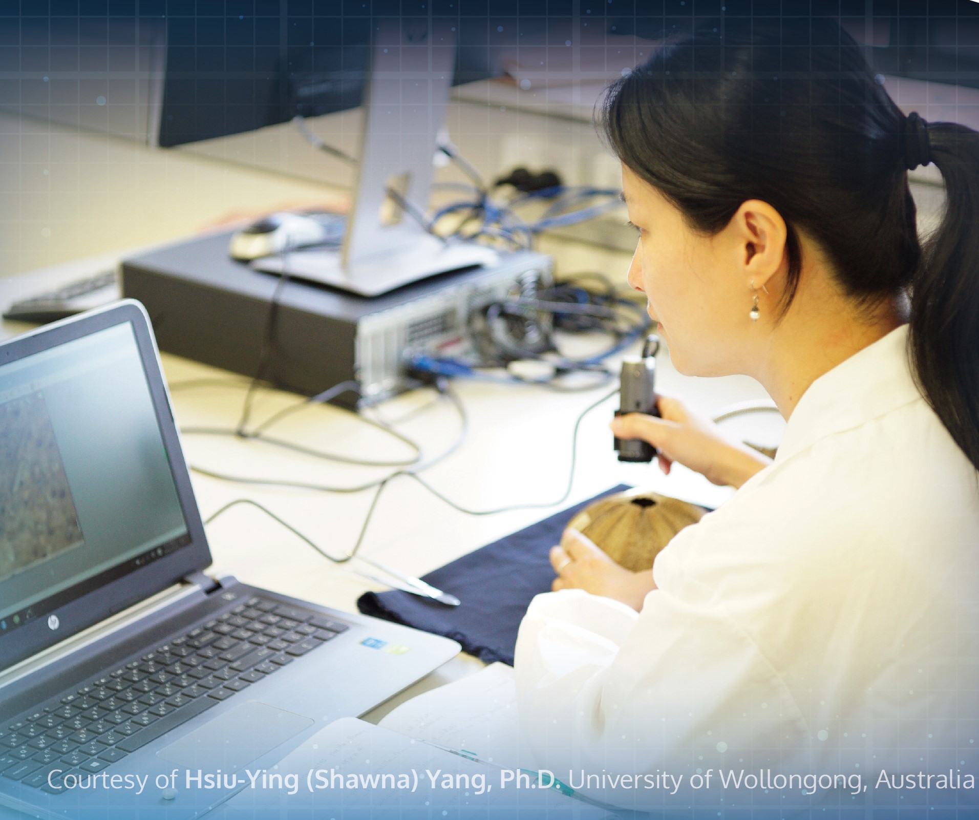
Dino-Lite digital microscope is a good support tool for art conservationists to restore and conserve objects and works of artists from the past. The portable digital microscope is held comfortably in your hand. It connects to a laptop via USB cable. The magnified image then appears on the screen through the DinoCapture 2.0 software. The alternative will be to look through an eyepiece of a stereo microscope. The Dino-Lite digital microscopes comes with high magnification and built-in LED lights to illuminate the object being examined. It is a portable microscope that will be a useful visual aid to see details that cannot be seen by the naked eye. The small and light form factor makes it useful for analysing large sized artworks which can be difficult to reach with the traditional stereo microscope.
When used to inspect sculptures, ceramics or architecture, the digital microscope can also help to identify the different types of stone, such as marble or limestone. It can also be used to analyse the components in mortars. With the DinoCapture 2.0 software, users will also be able to easily take pictures or videos for documentation. Other analysis tools available on the software includes measurements of line, circle, diameter, radius, angle, centre distance and etc. As such, it is a neat tool for measuring the size of the sand granules in mortar. It can also be used as a diagnostic tool for identifying the source of the stone’s deterioration, helping to identify salts, pollution and biological growth. It can even locate historic tool marks and past conservation treatments. When looking at a stain at a higher magnification, as a user, you would be able to differentiate if the stains come from pollutions or fungus for example.
The special lighting models with UV or infrared lights can help the user to view hidden details found on the art piece. For instance, the AM4115-FKT comes with 780 nm infrared LED lighting and records high quality images at 1.3 Megapixels. This model can also magnify objects up to 200x. It is ideal to be used to analyse chemical and physical properties of paper. On the other hand, the AF4515T-FVW comes with a 400 nm UV light source. The UV light source is a good aid for revealing historical moisture and mould damage to a paper. These are not always visible to the naked eye. Areas of the paper which were previously in contact with water, where size and the products which cause fluorescence have been removed, appear darker under UV light. All this information can then be used to aid the decision making process of forming a suitable conservation treatment.
Art restoration and conservation is a process that needs to be carried out very carefully. Art restorers would normally need to clean the art objects with great care so as not to damage the last original paint coat. They would need to differentiate between dirt, excess paint and original paint layer. They would also need to determine the authenticity of the items by looking at the object structure. With microscopes that can magnify items at 200x magnification, Dino-Lite can help to analyse in detail visually the different materials used to produce the art.
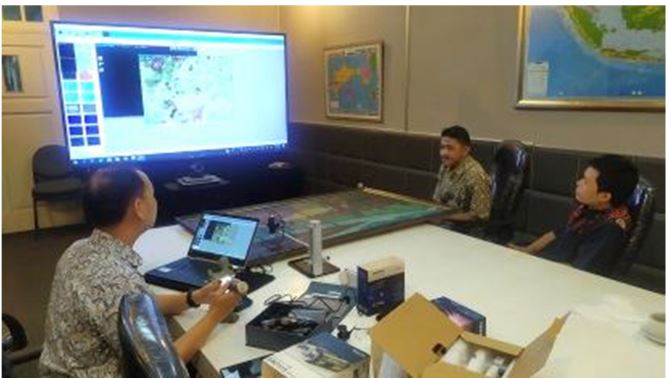
Dino-Lite being used to check a painting and projected on a wide TV screen.
The user will also find Dino-Lite USB microscope’s ability to capture images and videos, which can be annotated, to be very useful for documenting historical art objects. The user would also have the flexibility to select the medium they wish to view the art with. Dino-Lite digital microscopes can display images on TV screens, PC monitors, laptops and even Android devices. Art professionals can easily observe the work without any contact with the microscope’s ability to view items at a distance with the long working distance models of Dino-Lite.
Some might argue that without digital microscopes, conservators in the field can still turn to handheld loupes for analysis. However, the magnification powers of such loupes can only go up to about 30x. At the end of the day, the samples viewed will still need to be taken to a laboratory for analysis. Whereas the Dino-Lite USB microscope offers magnification of up to 250x. Analysis can be done immediately when you are on field. In the case of stone conservators, after looking at the stone with the Dino-Lite digital microscope, they can take samples, for example, of each kind of biological growth and bring them back to the lab for further analysis. The information can then be used to decide treatment plans for the long-term care and conservation of the stone monuments and buildings.
The Dino-Lite digital microscopes have time and again proven to itself to be a cost-effective tool. Coupled with the free DinoCapture 2.0 software, it will greatly help to speed up the analytical process in conservation studies of many different types of materials.
References
1. Nichols, E. (2016, December 14). Under the microscope – preliminary investigations of the changi archive conservation research project. Retrieved April 14, 2021, from https://specialcollections-blog.lib.cam.ac.uk/?p=13554
2. Lee, C. (2020, November 02). Conservation tools: The usb digital microscope. Retrieved April 14, 2021, from https://blogs.getty.edu/iris/conservation-tools-the-usb-digital-microscope/
Uses of Dino-lite Digital Microscope in Medical & Beauty
Practical platform for a routine check up and observation. Dino-Lite microscope provide one of the most important platform in studying biological sciences towards medical, trichoscopy and dermatology observation. It allows practitioner and patients to conveniently venture into the world of the cell, as well as discover the fascinating world of microscopic feature and draw a diagnosis precisely.
1. Dermatology - Scalp / Hair
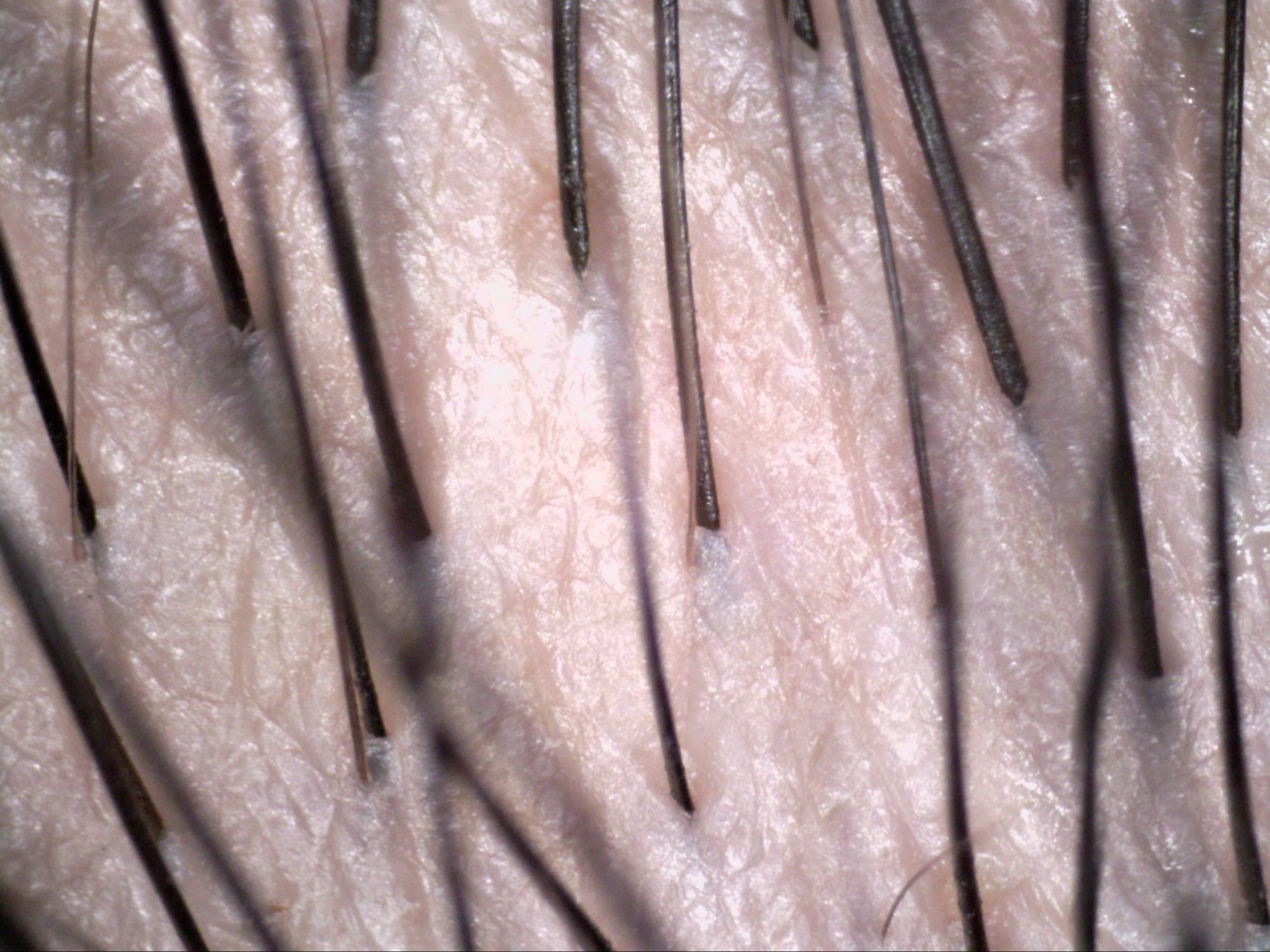
Here are some recommended Dino-Lite digital microscopes for dermatology-scalp/hair:
2. Nail Capillary
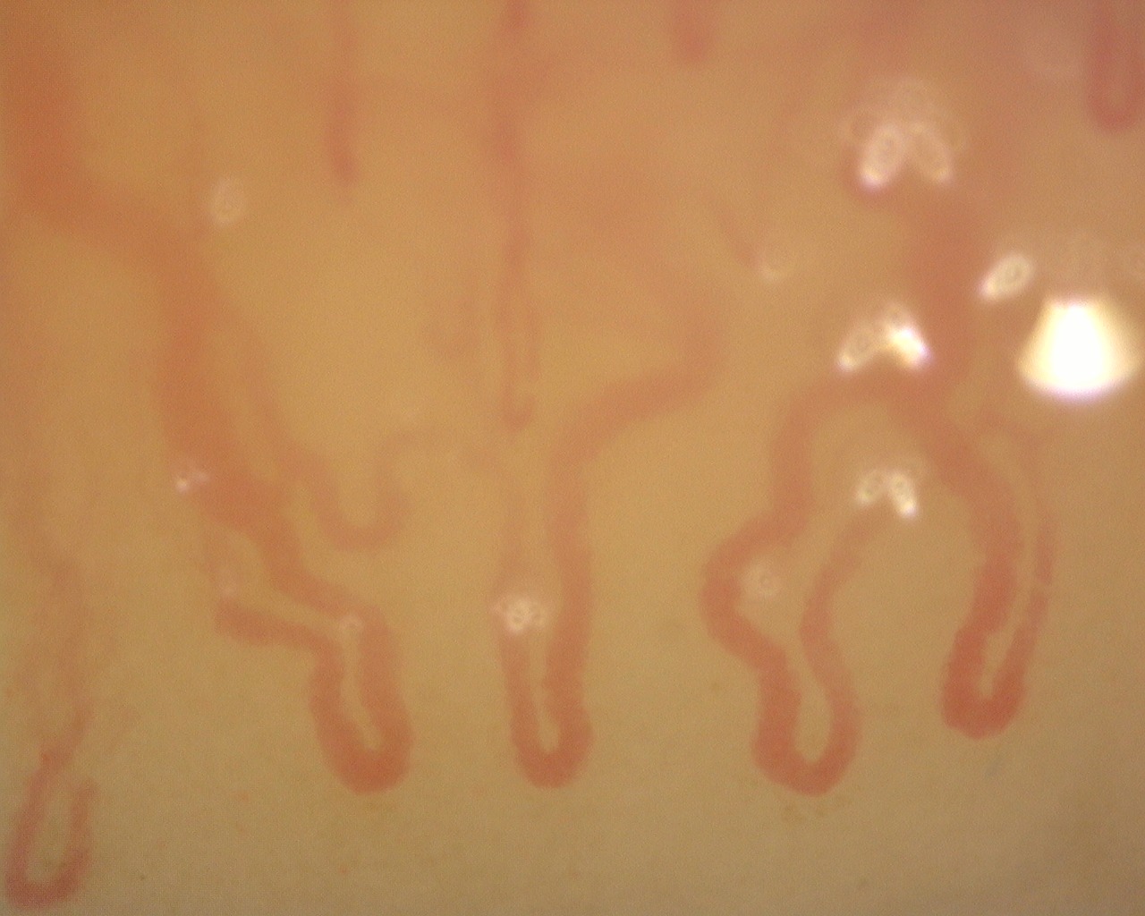
A capillary is a small blood vessel from 5-10 micrometres in diameter. They are the smallest blood vessels in the body: they convey blood between the arterioles and venules. Physician could diagnose various kinds pf disease by checking up the nail capillary.
Dino-Lite Nail Microcirculation Scope has a magnification of 500x and can examine the capillary with magnified clarity and digitally record the image for diagnostic and other purposes.
Here are some Dino-Lite Microscope recommended for observing nail capillary :
3. Ophthalmology

Ophthalmology is the field of medicine that deals with the anatomy, functions, and diseases of the eye. This includes the diagnosis and treatment of disorders of the eye. An ophthalmologist is a physician who specializes in ophthalmology. The credentials include a degree in medicine, followed by an additional four to five years of residency training in ophthalmology. Residency training programs for ophthalmology may require a one-year internship with training in internal medicine, pediatrics, or general surgery. Additional specialty training (or fellowship) may be sought in a particular aspect of eye pathology. Ophthalmologists are allowed to prescribe medications to treat eye diseases, implement laser therapy, and perform surgery when needed. Ophthalmologists may participate in academic research on the diagnosis and treatment of eye disorders.
Improved View
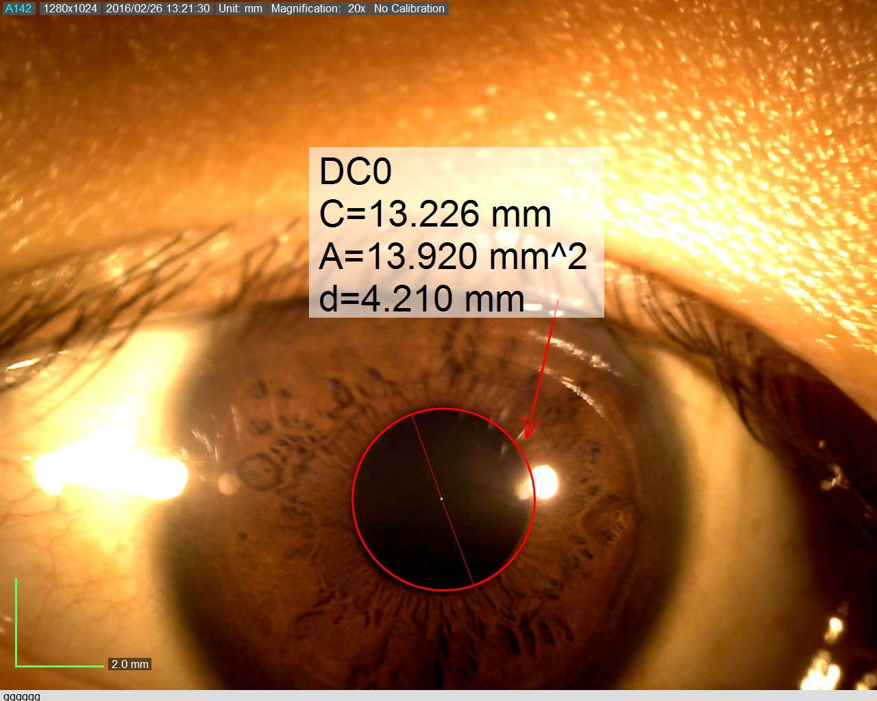
Dino-Lite digital microscopes can help ophthalmologists when you are doing iris examinations. The AM4113-RUT is one of the many models by Dino-Lite that is suitable for such work. It is a USB microscope which is used by connecting it to a computer. The main benefit of using digital microscopes is improved view. As a user, you would be able to examine the iris through a large screen monitor instead of hunching over patients. Ophthalmologists typically must hunch to get a close examination of the patients’ iris through oculars. The AM4113-RUT enables you to sit comfortably throughout the day as you are examining patients one after another. Extended periods of bending over oculars to examine multiple patients in a day may create a cumulative strain on your back. Over time, it can be damaging to your back and cervical spine. The traditional approach takes a lot of wear and tear on your bodies. Studies show that between 30% and 50% of ophthalmologist end up with cervical neck pain or herniated discs due to the way you must position your bodies to view the eye with a traditional microscope.
Ease of Use
The AM4113-RUT is easy to use. It is very much like the other Dino-Lite models where you merely use a single scroll to magnify and focus all at once. Your movements and hand placement may be different than using oculars. Most ophthalmologists are used to resting your head against the microscope as you look through the oculars. With the AM4113-RUT, you would no longer need to rest your head against the oculars of the microscope. It is a minor change that one can easily adapt to. Rest assured that the learning curve of using such a digital microscope is not steep. As a user, you would not require a long time to get accustomed to the Dino-Lite as it is as simple as viewing an image on the screen.
Data Collection
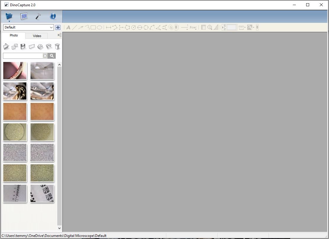
USB models of Dino-Lite digital microscopes are used with the DinoCapture 2.0 software. The application exists to support ease of documentation of your iris examination. You would be able to take pictures or videos by using the MicroTouch function. On the AM4113-RUT, there is a touch sensitive trigger for taking pictures and videos. Upon a single click, a picture is taken and automatically saved on your computer. All recent videos and images taken can be viewed on the left-hand bar of the DinoCapture 2.0 software. You can view the information seamlessly without interruptions during the examination. This is also a neat software for organizing and filing of patients’ iris examination images. It is easy to transfer images and videos from the DinoCapture 2.0 as they are saved to a designated folder in your computer. As you begin to use the digital microscope, you can get used to taking advantage of its capabilities. You will find an increase in efficiency as a benefit upon switching to the digital microscope. It will also greatly help you if you are seeking to file data in a “paperless” manner.
Communal Viewing
Anyone in the examination room — assistants, technicians, medical students — can watch the examinations on a monitor. As a result, it will help them to understand the patients’ situation better. If we compare it to the use oculars for examinations, only the person viewing through the microscope can get the complete picture. The others in the room will not be able to look at a monitor. The ability to see exactly what the doctor is seeing is an incredible advantage for teamwork overall. Everyone can better anticipate what is happening during the examination. Further, it also serves as a tool for resident education, fellow education, and medical student education because everyone is seeing the same thing at the same time.
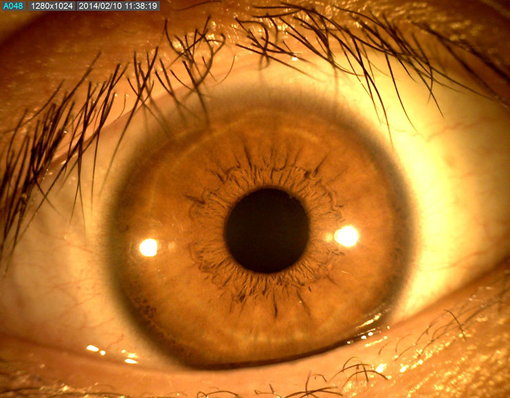
Increased Safety
In the COVID-19 pandemic, digital microscopes offer an additional barrier of safety for doctors and patients. Being able to examine patients through the monitors, and not through the oculars, allows the ophthalmologist to wear a full-face shield. There is a great safety advantage to having the ability to sit back and not hover directly over the patient’s face during examination. The ability to cover your face and eyes with a face shield is an important layer of protection, for both patients and doctors amid the pandemic. Information captured is also directly saved into the computer. You would not need to handle information on written copies. You would be able to access the images again by going through them in the computer. As a result, you would be less prone to error. Data that is captured goes straight from the digital microscope directly to the computer. It increases efficiency and is better for patient safety. When examining multiple patients, you would want to do it with as little room for error as possible. Digital microscopes close that gap quite a bit.
Here is the USB Dino-Lite Digital Microscope recommended for ophthalmology use:
Uses of Dino-lite Digital Microscope in Jewelry
Dino-Lite microscope provides one of the most important platforms in dealing with precious metals and gems. A jeweler put a price on precision, traceability, and investigative work which requires an concentration. Dino-Lite microscope allows jeweler and avid jewelry investor to conveniently venture into the microscopic world of precious metal, gems, as well as discover the fascinating value that sets them apart from the real deal and excellent replicas.
1. Diamond Examination
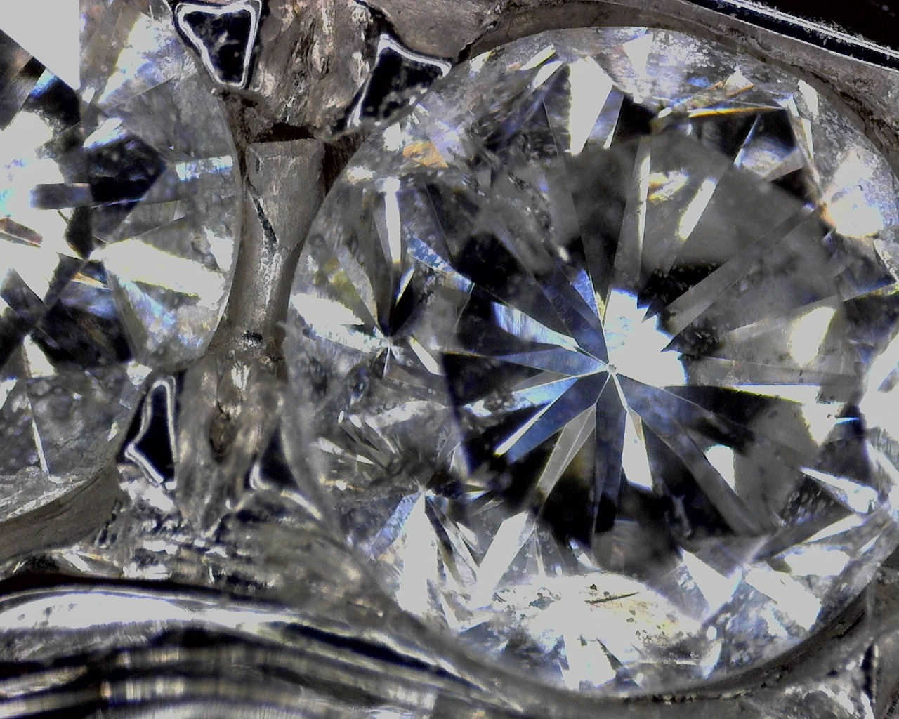
Diamonds certainly come in a variety of sizes, colors, and shapes. All diamonds possess intricate combinations of characteristics that make them unique. Most diamonds have minor inclusions and it is these internal markings that are very valuable in identifying your diamond, because there are no two diamonds that have inclusions in exactly the same places.
The other things that you look for are abrasions on the surface, nicks or cavities or cracks that may affect the structural integrity of the diamond. To examine diamond’s clarity, you will need some magnification tool. Dino-Lite Microscope for jewelers has the ability to examine stones, inspect jewelry for repairs, assist in appraisals and make imaging inventory fast and easy.
Here are some recommended Dino-Lite digital microscopes to examine diamonds:
2. Watchmaking / Repair
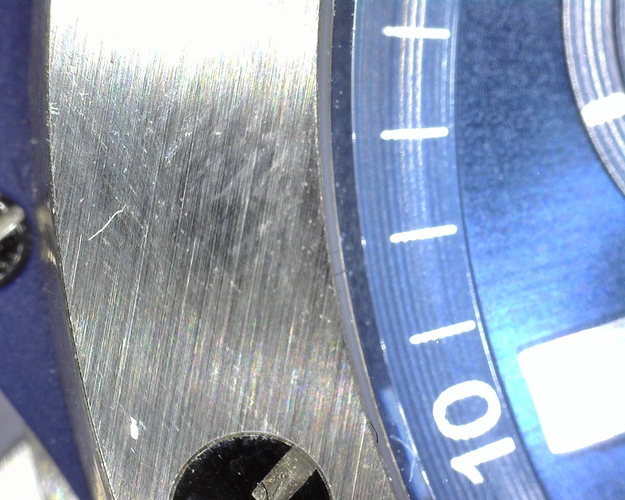
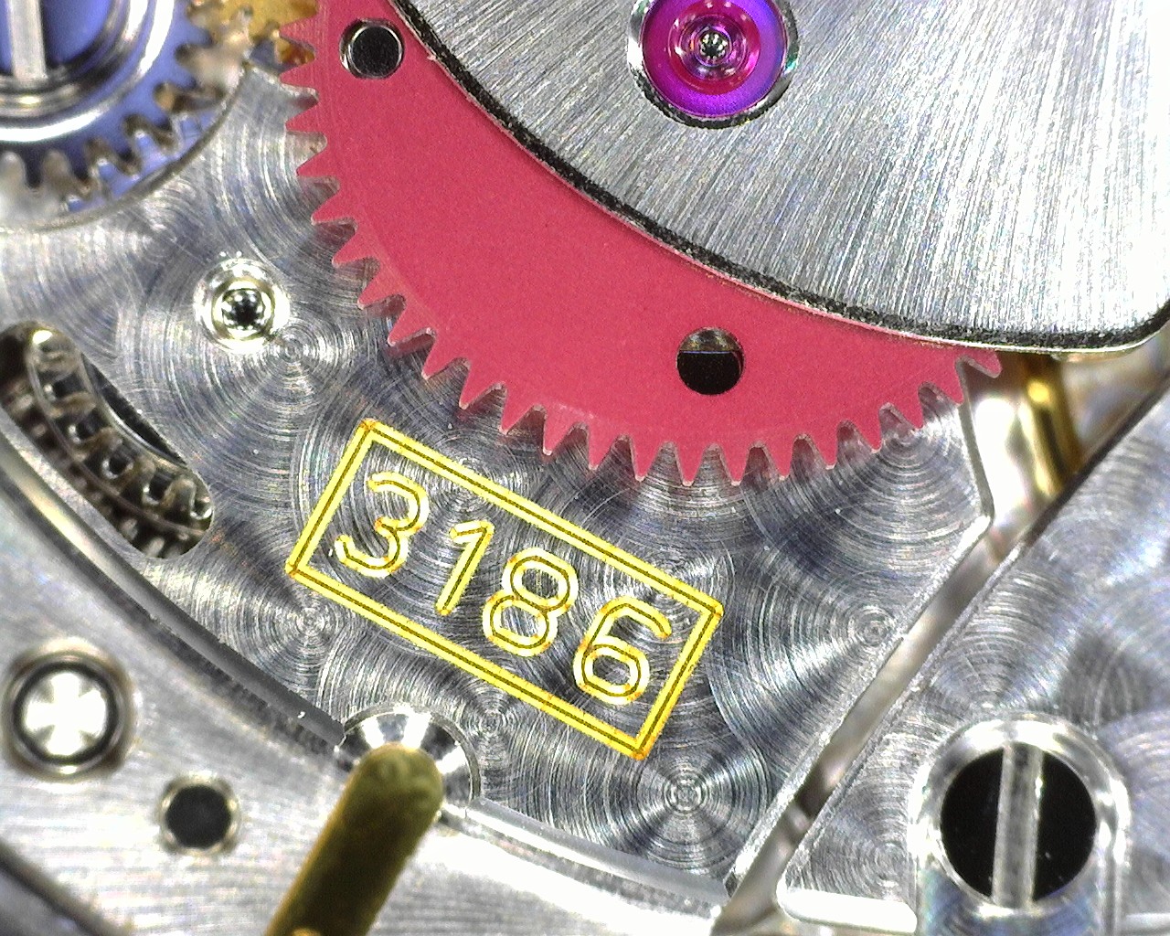
A good watch can consist of dozens, or even hundreds of mini parts. All parts need to be put together to make the watch work. Although most watches are now factory made, a watchmaker still needed to repair watches. Repairing watches can be a very intricate job to do, because of the number of tiny parts a watch has. Watchmaker will need to use magnifying tools that have polarized lighting. It helps provide uniform illumination to bring out more surface detail with minimal glare.
Here are some Dino-Lite Microscopes with polarizers recommended for watch making/repair:
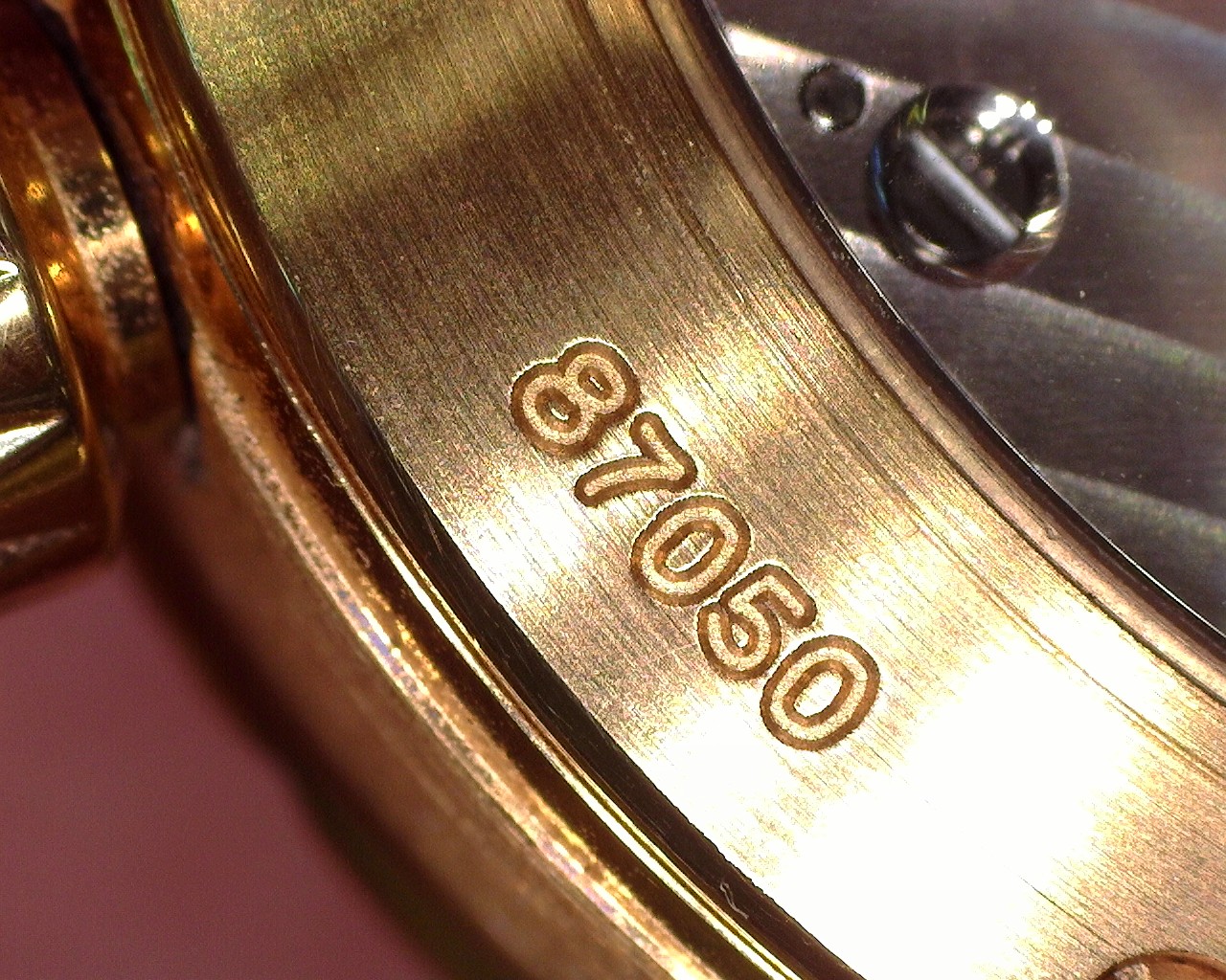
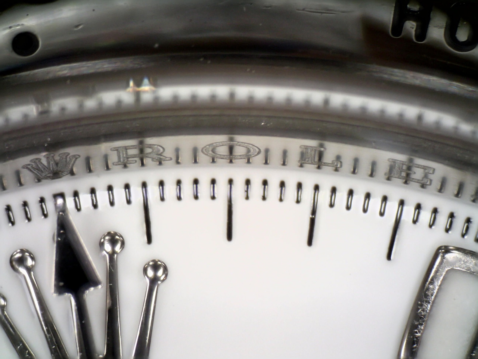
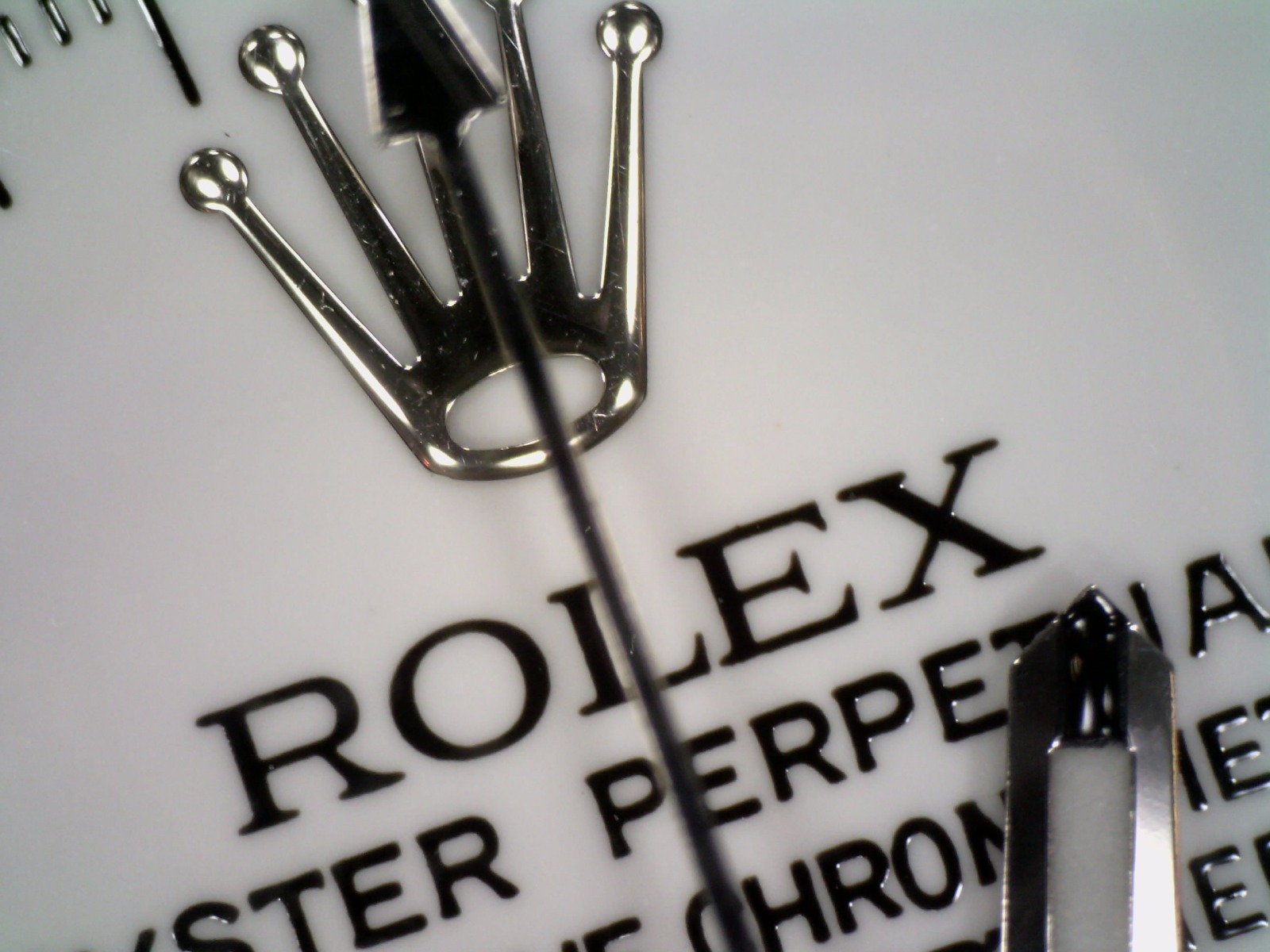
3. Watch Authentication
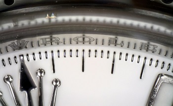
With a fast-growing tech-savvy population of watch enthusiasts, the purchase of luxury timepieces over the internet can be carried out with much ease and convenience. Any watch from any corner of the world could be purchased and couriered to your doorstep in a matter of days. Unfortunately, this ease and convenience has also led to a proliferation of replicas and outright fakes being spawned off as real deals, leading to substantial losses as a result.
Dino-Lite digital microscope is a handy and simple tool which can be used to ensure the provenance and authenticity of a watch. With the IP camera feature, you no longer need to take the trouble, time, and effort to personally travel to the source and personally inspect the watch. This can all be done remotely over the Internet. We are proud to be supporting digitalization of watch authentication today. To lessen the uncertainties, some watch brands have even developed ways to establish the identity of their watches by relying on blockchain, the database technology used for tamper-proof storage of digital information that is accessible to all. The high-quality images captured by Dino-Lite digital microscopes can be used for watch authenticators to verify the watch.
Uses of Dino-lite Digital Microscope in Industry
Dino-Lite is cleverly designed for industrial quality control engineers to perform visual inspections at the microscopic level. Many of the features will enable you to work with ease. The Dino-Lite models with the polarizer, for instance, is widely used in industrial inspections to reduce glare from highly reflective surfaces. The Dino-Lite microscopes with UV leds enables the user to detect minute scratches which cannot be easily detected by the naked eye.
Come talk to us and find the most suitable Dino-Lite digital microscope for you.
1. Textile - Fabric Inspection
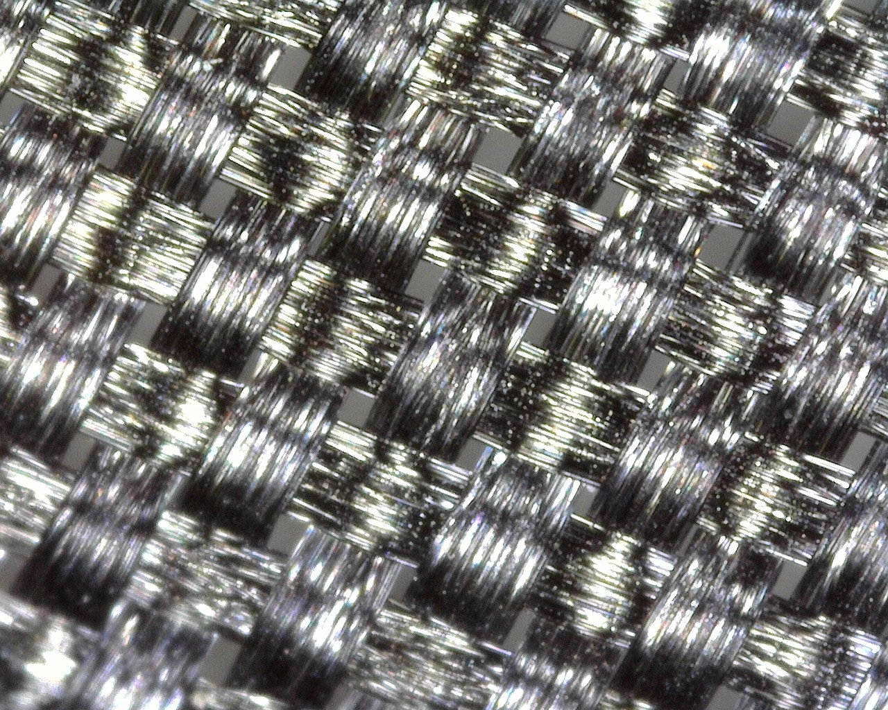
Dino-Lite digital microscope cameras allow Textile and Composite industry professionals the ability to see thread counts, contaminate section, and loom sewing monitoring with the ability to capture images and video with the included software.
2. Failure Analysis - PCB Board
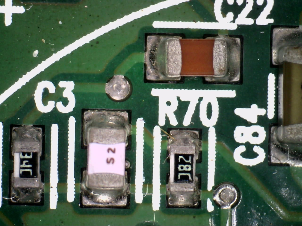
Failure analysis is the process of collecting and analyzing data to determine the cause of a failure, often with the goal of determining corrective actions or liability. The failure analysis process relies on collecting failed components for subsequent examination of the cause or causes of failure using a wide array of methods, especially microscopy.
Optical microscopy may be one of the most popular and preferred testing methods used for detecting faults, defects and problems associated with soldering and assembly. Many customers choose optical microscopy because of its speed and accuracy. Microscopy testing can verify improper construction, which can lead to stresses that can expose flaws at certain cross sections.
Dino-Lite has been used increasingly for failure analysis in the microelectronics industry, especially for printed circuit boards (PCBs). Digital microscopes are practical to use and allow an efficient workflow for inspection.
3. Identification of Genuine Currency Notes
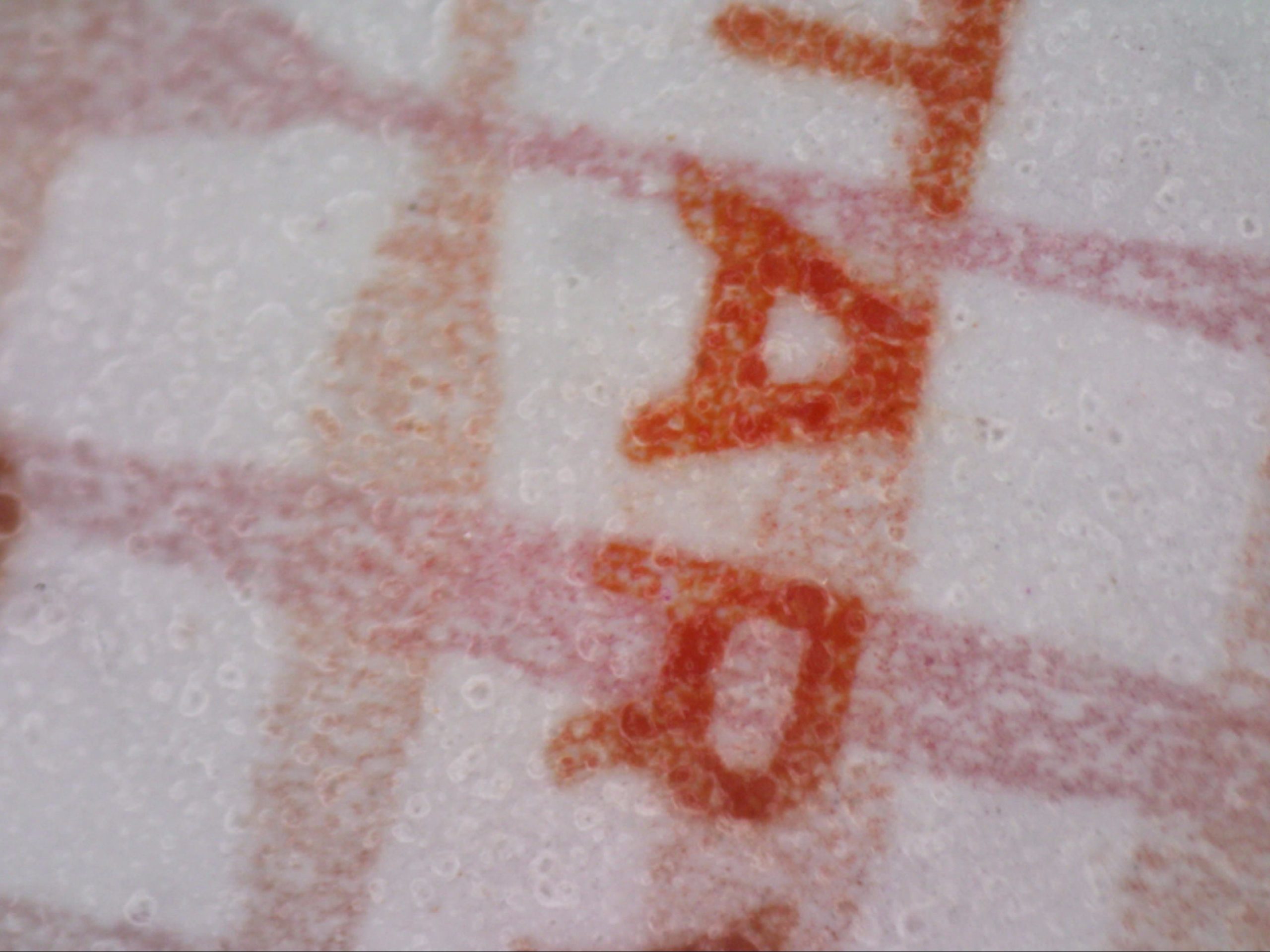
Microprinting is the production of recognizable patterns or characters in a printed medium at a scale that requires magnification to read with the naked eye. Micro texts security features are often added to currency notes and can be seen using the Dino-Lite digital microscope. Micro texts are added onto notes to identify genuine currency notes and also make it difficult to reproduce accurately using copiers or scanners. Typically, the text looks like a solid line at a distance.
Micro texts are used on currency, bank cheques, vouchers, and other items of value. It is often combined with other safety techniques such as holograms, void pantographs, prismatic printing, hot stamp foil printing, infrared visible inks or watermarks.
The portable nature of the Dino-Lite digital microscope makes it a very suitable tool for checking micro texts or prints on currency notes. You’d be able to bring it around. Coupled with the DinoCapture 2.0 software, it’s the perfect magnifier for checking currency notes and supporting report writing of suspicious currency notes.
4. Visual Inspection - Wire Bonding
5. Aerospace - Hairline Crack
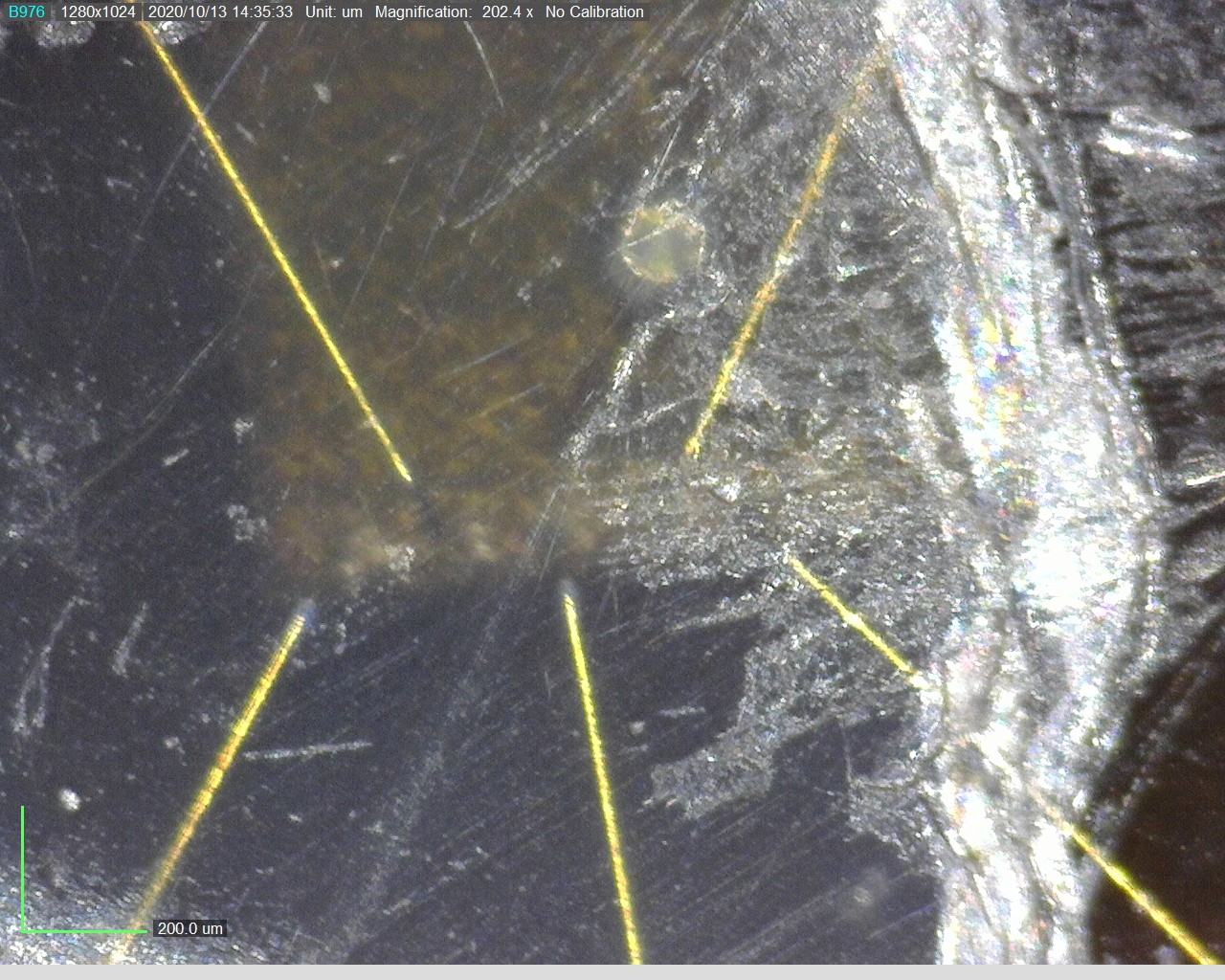
A crack is a type of discontinuity brought about by tensile stress, the result being that things are no longer held together. Hairline crack is a very thin crack that sometimes cannot be seen with naked eyes. In the aerospace field, hairline crack is not some mere problem that can be ignored. During flight, airplanes experience significant loads and stresses, and over time these loads can lead to cracks forming in high-stress areas of planes. If there is some hairline crack on the plane body, there’s a possibility that the plane will be unsafe to fly.
6. Gas Distribution Plate
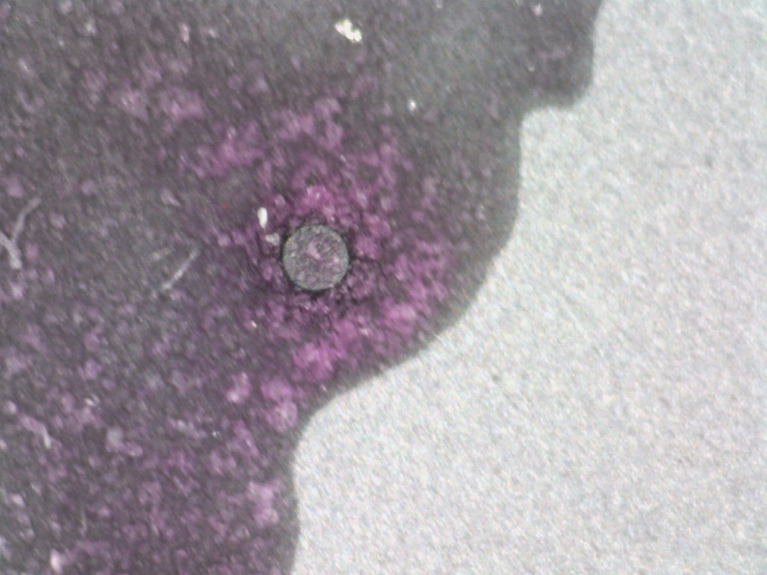
This Gas Distribution Plate is used in Semiconductor for electrodes, shower heads and Gas Distribution Plates. The Dino-Lite is used to see the small hole around the plate. We are checking to see if they are cleansed thoroughly with water. Normally, we would want to check this before and after cleansing.
7. Marking Inspection
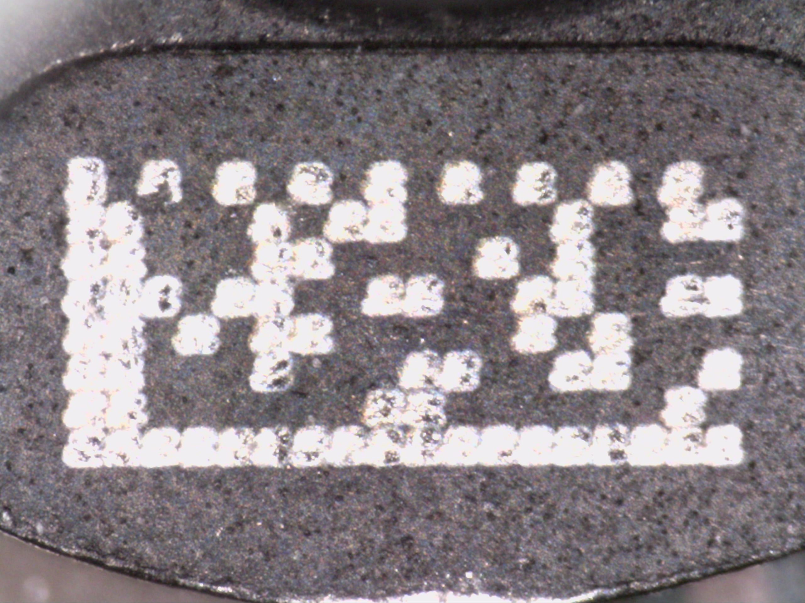
Dino-Lite is cleverly designed for industrial quality control engineers to perform visual inspections at the microscopic level. Many of the features will enable you to work with ease. The Dino-Lite models with the polarizer, for instance, is widely used in industrial inspections to reduce glare from highly reflective surfaces. The Dino-Lite microscopes with UV leds enables the user to detect minute scratches which cannot be easily detected by the naked eye.
Come talk to us and find the most suitable Dino-Lite digital microscope for you.

