
Microscopy has long been a cornerstone of scientific research, allowing us to explore the intricate details of the microscopic world. With technological advancements, microscopy has evolved significantly, providing researchers with powerful tools to observe and analyze samples with unprecedented clarity and precision. Among these tools, Dino-Lite stands out as a leader in the field, offering versatile and high-quality imaging solutions for a wide range of applications. In this article, we’ll explore some advanced imaging techniques with Dino-Lite and provide tips for achieving high-quality microscopy results.
Understanding Dino-Lite Microscopes
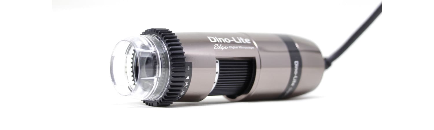
 Dino-Lite microscopes are renowned for their compact design, ease of use, and exceptional image quality. They utilize advanced optics and cutting-edge digital imaging technology to deliver clear, detailed images of microscopic specimens. Dino-Lite offers a variety of models tailored to different applications, including biological research, industrial inspection, forensics, and more. These microscopes feature adjustable magnification levels, adjustable LED illumination, and a range of accessories to enhance imaging capabilities.
Advanced Imaging Techniques with Dino-Lite
 In the realm of microscopy, Dino-Lite stands as a beacon of innovation and excellence, offering cutting-edge imaging solutions that empower researchers, educators, and enthusiasts to explore the microscopic world with unparalleled clarity and precision. With its compact design, user-friendly interface, and advanced imaging capabilities, Dino-Lite has revolutionized how we perceive and analyze the tiny wonders that lie beyond the threshold of the naked eye. In this section, we delve into advanced imaging techniques with Dino-Lite, uncovering the secrets of the microscopic universe through a lens of unprecedented sophistication and insight.
-
High-Resolution Imaging
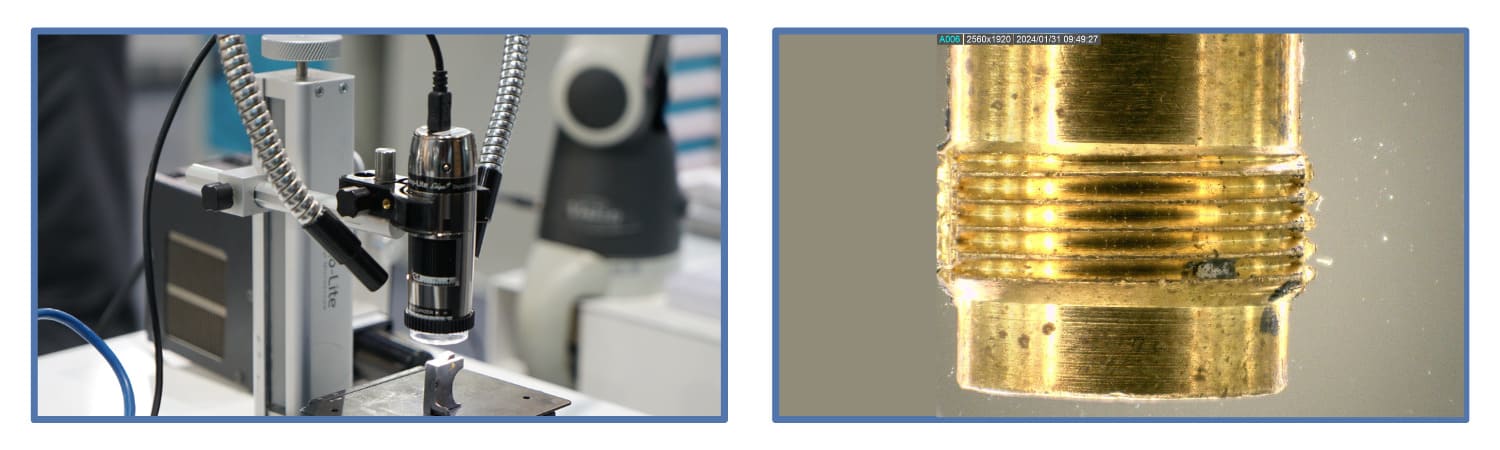
At the heart of Dino-Lite’s imaging prowess lies its ability to capture high-resolution images of microscopic specimens with breathtaking detail and fidelity. Equipped with state-of-the-art sensors and optics, Dino-Lite microscopes deliver images that rival the clarity and resolution of traditional laboratory-grade microscopes, allowing users to discern the subtlest features of their samples with precision and accuracy. Whether you’re examining biological specimens, geological formations, or industrial materials, Dino-Lite’s high-resolution imaging capabilities offer a window into a world of exquisite complexity and beauty.
-
3D Imaging and Depth Profiling
Beyond traditional 2D imaging, Dino-Lite microscopes can capture three-dimensional images of microscopic specimens, providing valuable insights into their topographical structure and morphology. By utilizing advanced imaging techniques such as structured light illumination and confocal microscopy, Dino-Lite enables users to visualize the three-dimensional architecture of their samples with unprecedented clarity and depth. From intricate cellular landscapes to rugged geological formations, 3D imaging with Dino-Lite unveils the hidden dimensions of the microscopic universe, offering a new perspective on the complicated interplay of form and function.
-
Fluorescence Imaging
Fluorescence microscopy is a powerful technique for visualizing specific molecules and structures within biological specimens, and Dino-Lite microscopes are equipped to harness their full potential. Dino-Lite enables users to visualize fluorescently labeled molecules and structures with exquisite specificity and sensitivity by incorporating fluorescence filters and light sources into their imaging systems. From tracking cellular processes’ dynamics to unraveling molecular interactions’ mysteries, fluorescence imaging with Dino-Lite opens new avenues for exploration in biological research and discovery.
-
Time-Lapse Imaging and Live Cell Imaging
Dynamic processes unfold in real-time within the microscopic world, and Dino-Lite microscopes can capture these phenomena through time-lapse imaging and live cell imaging. By capturing sequential images at defined intervals, Dino-Lite enables users to observe and analyze dynamic processes such as cell division, migration, and signaling with unparalleled temporal resolution. Whether you’re unraveling the mysteries of embryonic development or studying the behavior of living cells in response to external stimuli, time-lapse imaging with Dino-Lite provides a window into the dynamic rhythms of life at the microscopic scale.
-
Spectral Imaging and Hyperspectral Analysis
Spectral imaging is a powerful technique for analyzing the spectral characteristics of microscopic specimens, and Dino-Lite microscopes offer advanced capabilities in this domain. By capturing images at multiple wavelengths across the electromagnetic spectrum, Dino-Lite enables users to perform hyperspectral analysis, revealing valuable information about their samples’ chemical composition and molecular structure. Whether identifying specific biomolecules within cells or characterizing the composition of geological minerals, spectral imaging with Dino-Lite offers many opportunities for spectroscopic analysis and discovery.
Tips for High-Quality Microscopy with Dino-Lite
Microscopy, powered by Dino-Lite technology, opens a window to the intricate world of the microscopic, offering researchers and enthusiasts a glimpse into realms unseen by the naked eye. To harness the full potential of your Dino-Lite microscope and unlock the mysteries held within your samples, here are some advanced tips for achieving impeccable microscopy results:
-
Optimize Illumination
As mentioned earlier, proper illumination is the cornerstone of high-quality microscopy. With Dino-Lite microscopes, fine-tune the LED illumination to match the specific characteristics of your sample. Avoid extreme overexposure or underexposure, as these can obscure details and compromise contrast, leading to subpar images that fail to do justice to your samples’ intricacies.
-
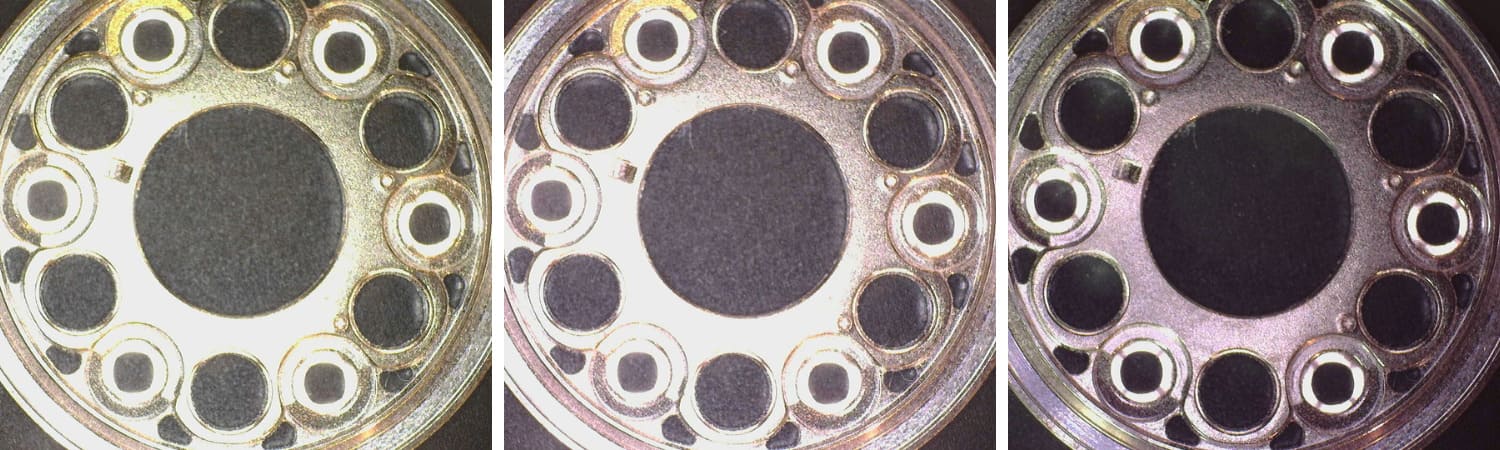
Use Polarization
 Harness the power of polarization many Dino-Lite models offer to combat glare and elevate contrast in your images. By experimenting with different polarization settings, you can minimize unwanted reflections and unveil the delicate nuances hidden within transparent or reflective samples, revealing a wealth of detail that might otherwise go unnoticed.
-
Adjust Focus and Magnification
 Precision is paramount when it comes to focus in microscopy. Utilize the fine focus knob on your Dino-Lite microscope to meticulously adjust focus, ensuring that your images are sharp and crisp. Furthermore, explore the range of magnification levels available to capture every intricate detail of your samples, from the grandeur of macroscopic structures to the subtleties of cellular morphology.
-
Explore Different Lighting Modes
 Dino-Lite microscopes offer many lighting modes, each tailored to illuminate specific types of samples optimally. From traditional brightfield illumination to the dramatic contrasts of darkfield and the nuanced revelations of differential interference contrast (DIC), experiment with different lighting modes to uncover the full spectrum of details in your samples, choosing the mode that best complements your specific application.
-
Utilize Measurement and Annotation Tools
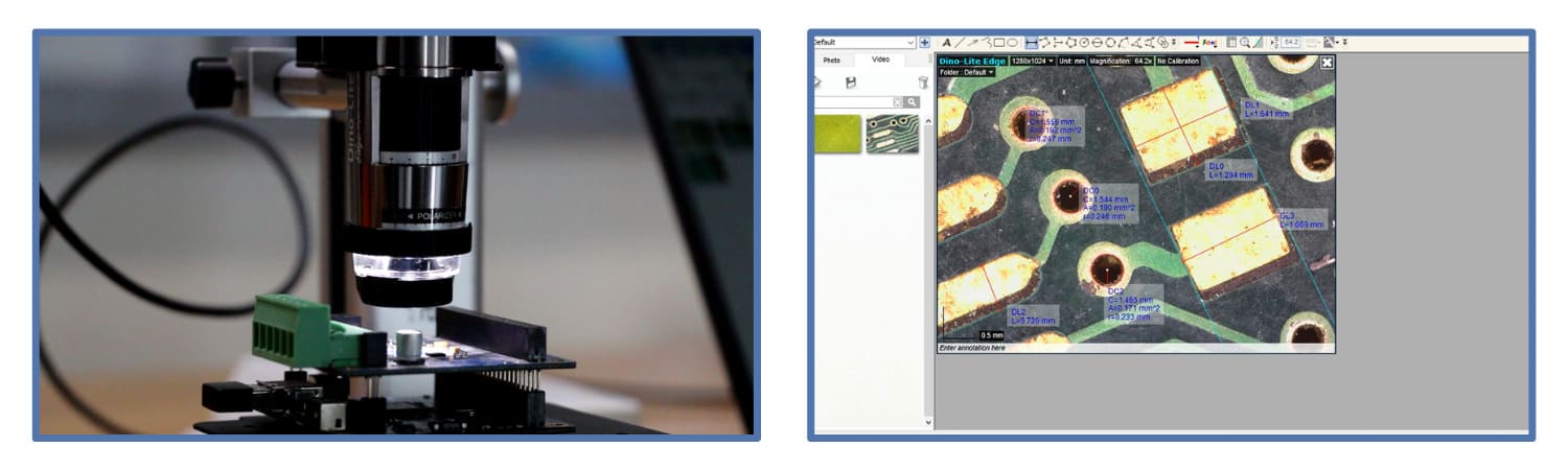 Beyond mere observation, leverage Dino-Lite software’s measurement and annotation tools to delve deeper into your microscopy findings. Accurately measure dimensions, annotate vital features, and generate comprehensive reports for further analysis and sharing, transforming your microscopy endeavors into a rich tapestry of quantifiable data and insightful observations.
-
Calibrate Your Microscope
Adhere to regular calibration procedures for your Dino-Lite microscope to maintain the integrity of your microscopy data. Follow the manufacturer’s guidelines meticulously, conducting calibration checks at regular intervals to ensure precise measurements and consistent imaging results, laying a solid foundation for your scientific endeavors.
-
Consider Environmental Factors
Environmental conditions can significantly impact microscope performance and image quality. To mitigate potential disruptions, ensure that your Dino-Lite microscope is in a stable environment with controlled temperature, humidity, and minimal vibrations, providing a conducive setting for optimal imaging results.
-
Practice Proper Sample Preparation
Last but certainly not least, meticulous sample preparation is the bedrock upon which high-quality microscopy is built. Adhere to established protocols for sample fixation, staining, and mounting, ensuring that your samples are meticulously prepared to withstand the scrutiny of Dino-Lite microscopy. Investing time and effort into proper sample preparation will pave the way for insightful observations and groundbreaking discoveries.
